
Clique nas imagens para assistir aos vídeos.
Neurinoma do Acústico
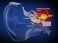
This growth is a benign tumor that forms on the vestibulocochlear nerve. This nerve leads from the inner ear to the brain. Acoustic neuromas usually grow slowly and do not spread. However, they can eventually grow so large that they press against surrounding structures, including the brain and other nerves.
Tumor da Hipófise
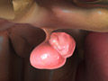
Your pituitary gland is found just under your brain. This pea-sized gland makes hormones that affect many of your body’s functions. A pituitary tumor can cause it to release too much or too little of these hormones. This can cause serious problems.
Anatomia do Cérebro

The brain is the control center of the human body. It forms your thoughts and preserves your memories. It regulates your body’s actions, from the movements you choose to perform to the functions you don’t even consciously think about. Let’s take a closer look at the anatomy and the function of the brain.
Astrocytoma

This is a tumor that begins in a brain cell called an “astrocyte.” These cells help give your brain its structure. An astrocytoma can form in your brain, in your brain stem or in your spinal cord. There are many types of astrocytomas. They can be cancerous or noncancerous. They can grow slowly or quickly. A doctor can figure out the specific type you have.
Aneurisma Cerebral
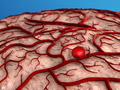
This condition is a bulge that forms in the wall of a weakened artery in the brain. This bulge can leak or rupture, causing a stroke. An aneurysm can be life-threatening.
Tumor Cerebral

This is a mass of abnormal cells. It may be inside your brain, or it may be next to your brain. It can grow and press harmfully against healthy brain tissue. This can cause a wide range of problems throughout your body. A brain tumor can severely impact your life.
Cerebral Cavernous Malformation (CCM)

This is a mass of enlarged blood vessels in your brain or spinal cord. Pockets in the mass slow down or even trap blood. This can lead to blood clots, or to a leaking of blood we call a “hemorrhage.”
Hematoma Subdural Crônico (Hemorragia)

This condition is a buildup of clotted blood between the brain’s outer layer and the membrane that covers the brain (called the dura). It usually occurs in the elderly, and can be caused by even a minor bump to the head.
Epilesia

This is a problem with the electrical activity of your brain’s nerve cells. These cells are called “neurons.” With epilepsy, they sometimes send out disorganized signals. When this happens, you can suddenly lose control of your body for a brief time. There may be a change in how you act or feel. We call this a “seizure.”
Hidrocefalia

This condition is caused by an increased amount of cerebrospinal fluid (commonly called CSF) in the brain’s ventricles. The ventricles are a system of large, fluid-filled open spaces inside the brain. Too much CSF in the ventricles can elevate pressure in the skull. It can damage delicate brain tissue.
Meningioma

This is a tumor in your meninges. These thin layers of protective tissue surround your brain and spinal cord. Most meningiomas are not cancerous. They usually grow slowly.
Tumor Cerebral Metastático

This is a cancer that began elsewhere in your body and then spread to your brain, forming one or more tumors. Many different cancers can spread this way. These tumors are actually more common than tumors that begin in the brain’s own tissues.
Hidrocefalia de Pressão Normal (NPH)

This condition, which usually occurs in adults 55 and older, is an excessive accumulation of cerebrospinal fluid (CSF) in the ventricles of the brain. The ventricles are a system of large, fluid-filled open spaces inside the brain. Too much CSF in the ventricles can distort the brain’s shape. It can make the brain susceptible to injury.
Convulsão

This is a sudden burst of electrical activity in your brain. It overwhelms parts of your brain, usually for no more than a few minutes. Most seizures don’t cause lasting harm.
Apreensão do Lobo Temporal

This type of seizure begins in one of the temporal lobes of the brain. It happens because of abnormal electrical activity. Temporal lobe seizures can severely impact your daily life.
Surgical Care and Management
Clipping de Aneurisma
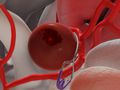
This surgical procedure is performed to treat an aneurysm, a bulge in the wall of an artery, inside the skull. Aneurysms can often become so large that they rupture or leak. In this procedure, a small, metal clip is applied to the base of the aneurysm to prevent blood leakage.
Craniectomy for Chiari Malformation (Foramen Magnum Decompression)
This surgery is used to treat Chiari malformation, an abnormality that results in a part of the brain extending into the upper spinal canal. During the procedure, small sections of bone are removed from the rear of the skull and spine to create more space for the errant brain tissue.
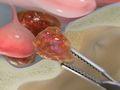
Craniotomy for Meningioma

This surgery removes a tumor called a “meningioma.” That’s a type of tumor that begins in the thin tissue that surrounds your brain and spinal cord.
Craniotomy for Tumor

This procedure, performed under general anesthesia, creates an opening through the skull for brain tumor removal. The surgery usually requires between two to five hours to complete. The length of surgery depends on the type and size of the tumor.
Ommaya Reservoir Placement
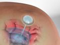
During this procedure, the surgeon places a small dome-shaped reservoir beneath the scalp and connects it to a fluid-filled cavity in the brain. Once in position, the ommaya reservoir can be used to administer medications or to withdraw fluid.
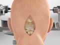
Cirurgia de tumor hipofisário (abordagem transesfenoidal)
This surgery treats one or more tumors on or near your pituitary gland. That’s a small organ at the base of your brain. Your surgeon will reach the pituitary gland through your nostrils.
Biopsia Cerebral Estereotáxica (Método de Biópsia por Agulha)
This is a way for a surgeon to take a sample of abnormal tissue from inside your brain. It’s done with a needle that’s carefully guided into your brain.
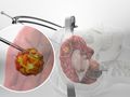
Suboccipital Craniectomy for Acoustic Neuroma
This surgery is used to remove an acoustic neuroma, a type of noncancerous tumor that forms on a nerve in the middle ear. The procedure is performed under general anesthesia and requires a hospital stay.
Ventriculoperitoneal Shunt for Hydrocephalus

During this surgical procedure, a small drainage tube is implanted to relieve the pressure of hydrocephalus. Hydrocephalus is a condition that develops when excess cerebrospinal fluid builds up within the ventricles of the brain.
Peripheral
Non-Surgical Care and Management
Monitoramento Neurofisiológico Intraoperatório (IONM; IOM)

This is a way to monitor your nerves during surgery. It gives your surgical team real-time feedback. It helps keep your nerves safe during your procedure. Here’s how it works.